Zhang Huazheng1,
Wang Penglong2,
Ren Liwei1,
Wang Xiaobo2,
Li Guoliang2,
Wang Mina1,
Chu Fuhao2,
Gong Yan2,
Xu Bing2,
Bi Siling1,
Lei Haimin2,
Zhang Yuzhong1 ![]()
For correspondence:- Zhang Yuzhong Email: hm_lei@126.com Tel:+861064286964
Received: 4 August 2015 Accepted: 29 December 2015 Published: 28 February 2016
Citation:
Huazheng Z, Penglong W, Liwei R, Xiaobo W, Guoliang L, Mina W, et al.
Proliferative activity and neuroprotective effect of ligustrazene derivative by irritation of vascular endothelial growth factor ex
© 2016 The authors.
This is an Open Access article that uses a funding model which does not charge readers or their institutions for access and distributed under the terms of the Creative Commons Attribution License (http://creativecommons.org/licenses/by/4.0) and the Budapest Open Access Initiative (http://www.budapestopenaccessinitiative.org/read), which permit unrestricted use, distribution, and reproduction in any medium, provided the original work is properly credited..
Purpose: To investigate the proliferative activity and neuroprotective effect of a newly identified ligustrazine derivative (4-((3,5,6-trimethylpyrazine-2-yl)methoxyl)-3-methox-ybenzoic acid-3,5,6-trimethylpyrazin- 2-methyl ester, T-VA) and the possible mechanism related to vascular endothelial growth factor (VEGF) in cerebral ischemic injury.
Methods: The pharmacological activity of T-VA was evaluated using MTT ((3-(4,5-dimethylthiazolyl2-yl)-2,5-diphenyltetrazolium bromide)) assay, while cellular morphology was observed with hematoxylin and eosin (HE) staining. Chick chorioallantoic membrane (CAM) model, immuno-histochemical analysis, and enzyme-linked immunosorbent assay (ELISA) were used to determine the ex
Results: T-VA promoted neuron activity, and the doses of 15 and 30 μM showed more significant effect (p < 0.05). The viability of PC12 cells increased significantly in T-VA (30 and 60 μM) groups (p < 0.05) and increased in a dose-dependent manner. Immunohistochemical analysis showed stimulated VEGF ex
Conclusion: These findings indicate that T-VA helps to prevent ischemic injury by increasing VEGF ex
Introduction
Ischemic stroke has attracted attention in domestic and international research, and several reviews have recently been published about the effect and potential benefits of traditional Chinese medicine in the treatment of cerebral ischemic injury [1]. It has been suggested that some herbal medicines, or their products, may have the functions of improving microcirculation in the brain, protecting against ischemic injury, improving cerebrovascular properties, and inhibiting neuron apoptosis [2-4]. The lack of effective and widely applicable pharmacological treatments for ischemic stroke patients may explain the growing interest in discovering drugs derived from traditional medicines [5].
As one of the major effective ingredients of Rhizoma Chuanxiong ligustrazine (2,3,5,6-tetramethylpyrazine, TMP) has been widely used in the treatment of ischemic stroke and cerebrovascular disease in China for many years [6] Previous studies have shown various pharmacological activities of TMP, such as anti-myocardial ischemia, anti-cerebral ischemia, inhibiting platelet aggregation, protecting vascular endothelial, etc [7-11]. To further improve the neuroprotective property of TMP, we decided to undertake a study of a novel series of TMP derivatives based on the principles of hybridization and prodrug design in medicinal chemistry and acquired compounds with pharmacologically additive or synergetic effects [12-14]. It has been demonstrated that T-VA ((4-((3,5,6-trimethylpyrazine-2-yl)methoxyl)-3-methoxybenzoic acid-3,5,6-trimethylpyrazin- 2-methyl ester; see Results, A) has a promising protective effect against CoCl2-induced neurotoxicity in differentiated PC12 cells (EC50 = 4.249 µM) [15] and that the plasma concentration of T-VA reaches peak levels between 4 and 6 h post-intragastric administration [16]. Interestingly, some studies show that congener structures of T-VA possess protective effects on damaged vascular endothelial cells, such as ECV-304 cells, human umbilical vein endothelial cells (HUVECs) and Hy-926 cells [17-19]. Moreover, ligustrazine derivatives showed good antiplatelet aggregation activity [19].
Based on the aforementioned evidence, we reasoned that the neuroprotective effect of T-VA might be associated with its thrombolytic and vascular biological properties. Vascular endothelial growth factor (VEGF), an angiogenic protein, is able to enhance survival ability of endothelial cells and has a role in thrombus resolution that was helpful in the treatment of cerebral ischemia [20,21]. This potential therapeutic value and neuroprotective effect in ischemia might be mediated by VEGF itself or by a signal transduction pathway activated by VEGF [22]. VEGF was shown to attenuate acute neuronal death and play a neuroprotective role under oxygen-glucose deprivation conditions through VEGFR-2 [23]. Moreover, when VEGF was used to treat focal cerebral ischemia in rats, the infarct volume was reduced and neurological functions were improved [24]. Therefore, our aim was to investigate the proliferative activity and neuroprotective effect of T-VA and the possible mechanism related to VEGF in cerebral ischemic injury.
Methods
General
Rats (female rats pregnant for 16-18 days and male SD rats weighing 280-300 g) were purchased from Beijing Vital River Laboratory Animal Technology Company Limited (Beijing, China) and kept under standard laboratory conditions (tap water, constant room temperature, 22 °C). All animal protocols were approved by the Ethics Committee of Beijing University of Chinese Medicine in accordance with the National Institutes of Health guidelines (Contract 2012-0036, June 2012). All efforts were made to minimize animal suffering and to reduce the number of animals used. PC12 cell line was purchased from the Institute of Materia Medica of the Chinese Academy of Medical Science. Fertilized White Leghorn chicken eggs were provided by Chinese Academy of Agricultural Sciences.
Animals, neurons culture and drugs
Fetal rats were taken from pregnant rats and washed twice with 4 °C phosphate buffered saline (PBS). After stripping the meninges and vessels, the brains of fetal rats were cut into 1 mm3 cubes. All steps were conducted on ice. Neurons were isolated by 0.1 % trypsin digestion for 10 min in a 37 °C incubator and fixed with Dulbecco’s modified eagle medium supple-mented with 10 %(v/v) fetal bovine serum and 100 U/mL penicillin-streptomycin (Thermo Technologies, New York, NY, USA) [25,26]. Then neurons were seeded onto poly-L-lysine-coated 96-well culture plates at 8 × 10⁴ cells/well, and the plates were incubated at 37 °C in a humidified atmosphere of 5 % CO2 for 48 h. Subsequently, the neurons were treated with different doses of T-VA (60, 30, and 15 μM) and nimodipine (NMDP) for 24 h.
PC12 cell culture and drugs
PC12 cells were cultured in RPMI 1640 medium supplemented with 5 % (v/v) fetal bovine serum, 10 % (v/v) horse serum and 100 U/mL penicillin-streptomycin (Thermo Technologies, New York, NY, USA) at 37 °C in a humidified atmosphere of 5 % CO2. When cells achieved the desired density of > 80 % confluency, the original medium was removed and cells were cultured with a serum-free medium for 12-14 h. Then the cells were suspended in the RPMI 1640 medium supplemented with 10 % (v/v) fetal bovine serum, and seeded into poly-L-lysine-coated 96-well culture plates at 7 × 10⁴ cells/well, differentiated by treated with nerve growth factor (NGF; 50 ng/mL) for 48 h [2]. Subsequently, the differentiated PC12 cells were treated for 36 h with various concentrations (60, 30, 15 μM) of T-VA or RPMI 1640 medium supplemented with 10 % (v/v) fetal bovine serum. T-VA was dissolved in dimethyl sulfoxide (DMSO). The final concentration of DMSO was < 0.1 % (v/v).
MTT (3-(4,5-dimethylthiazolyl2-yl)-2,5-diphenyltetrazolium bromide) assay
After MTT solution (20 μL, 5 mg/mL) was added to each well, the plate was incubated for an additional 4 h at 37 °C. The supernatant was removed carefully by pipetting from wells without disturbing the attached cells. Formazan crystals were solubilized by adding 150 μL of DMSO to each well, and then the plate was shaken for 15 min [2]. The absorbance of neurons at 540 nm and PC12 cells at 490 nm were measured with a Bio-Rad 550 spectrophotometer (Bio-rad, California, CA, USA).
Hematoxylin and eosin (HE) staining method
After pretreatment with the serum-free medium for 12-14 h, cells were seeded at a concentration of 7 × 10⁴ cell/mL on a poly-L-lysine-coated sterile cover slip in 24-well tissue culture plates, and differentiated by treatment with NGF (50 ng/mL) for 48 h. The differentiated PC12 cells were pretreated with T-VA for 36 h at the optimum concentration (60 μM), which was selected by MTT assay. Cover slips were washed twice with PBS, removed and treated with acetone for 10 min at -20 °C. Then cells were washed with PBS and stained with hematoxylin for 3 min. The stained nuclei were rinsed with running water for 1 h followed by eosin staining for another 2 min. Finally, stained sections were dehydrated using gradient ethanol and cleared in xylene [2,27]. The cellular morphology was photographed by light microscopy (Nikon, Kobe, Japan).
Immunohistochemical analysis of VEGF
Our previous study demonstrated that T-VA protect CoCl2-induced neurotoxicity in differentiated PC12 cells [2], so all measurements were performed after PC12 cells were induced by CoCl2 (dissolved in RPMI 1640 medium, 200 mM) for 12 h. Cells in the NGF group were not treated with T-VA and CoCl2. Briefly, after being washed three times with PBS, PC12 cells were fixed in acetone for 10 min at -20 °C. After being blocked in 3 % H2O2-CH3OH for 30 min at 37 °C, the cells were incubated with VEGF antibodies overnight at 4 °C. Cells were then incubated in horseradish peroxidase-conjugated AffiniPure goat anti-rabbit immunoglobulin G (IgG) diluted to 1:250 in PBS for 30 min at 37 °C, followed by streptavidin-biotin complex (SABC) for 30 min at 37 °C. 3, 3-Diaminobenzidine (DAB) was used as a chromogen for 10 min. PBS was used between each step to wash the cover slips. Finally, PC12 cells were stained with hematoxylin for 30-60 s and washed with running water for 1 h. Stained sections were dehydrated using gradient ethanol and cleared in xylene. The expression of VEGF was observed and photographed under a microscope [28].
Chick chorioallantoic membrane (CAM) model
The eggs were placed in an incubator immediately after embryogenesis started and were kept under constant humidity of 65 % at 37 °C. On the 7th day, a square window was opened on the shell and physiological saline (0.1 mL) was injected in to detach the shell membrane. T-VA was carried by gelatin sponges and implanted at 10 and 20 μg/egg. The control group was treated with physiological saline. The windows were sealed with medical adhesive tape. The aforementioned treatment steps were all performed under sterile conditions. On the 11th day, the tape was removed and the entire CAM was detached after tissue fixation with methanol/acetone (1:1, v/v). Then, computer-assisted image tracking was used to obtain absolute values for the number of microvessels that were 1-100 μm in diameter [15]. The model was established according to previously published method [29] and our previous studies [30].
Middle cerebral artery occlusion (MCAO) model
Transient MCAO was conducted in male rats according to the previously published methods [31,32]. There were 15 rats in each of the MCAO and sham-operated groups. Briefly, male rats were anesthetized with 10 % chloral hydrate (350 mg/kg), and the right common carotid artery and external carotid arteries were isolated and ligated. A nylon suture (diameter 0.25 mm) was advanced from the common carotid artery to the internal carotid artery for a distance of approximately 18-20 mm from the external-internal carotid artery. The blocking of the middle cerebral artery induced cerebral ischemia. Then the muscle and skin were sutured with 4-0 nylon. Penicillin (4 million units/mL) was given to the rats for 3 days following the operation. In sham-operated rats, internal and external carotid arteries were also exposed and separated. When the rats recovered consciousness, 6 h after the operation, scores were graded as follows: according to Bederson’s description [33]: 0, no deficit; 1, failure to fully extend left forepaw; 2, circling to the left; 3, falling to the left; 4, no spontaneous walking with a depressed level of consciousness. Rats with scores from 1 to 3 were selected for further testing and were divided randomly to the ischemia group, T-VA group (120 mg/kg and 60 mg/kg), and NMDP group. The rats were treated with T-VA by intragastric administration (1 mL/100g) once a day until the 10th day. The sham and model groups were treated with isovolumetric 0.5 % sodium carboxymethyl cellulose (CMC-Na).
Enzyme-linked immunosorbent assay (ELISA) for VEGF detection
The level of VEGF in serum was measured with sandwich ELISA kits. Optical densities were determined at 450 nm with a Bio-Rad 550 spectrophotometer.
Statistical analysis
Statistical comparisons between groups were made by one-way ANOVA followed by Student’s t-test using the SPSS version 20. Results were considered significant at p < 0.05.
Results
Effect of T-VA on neuron activity
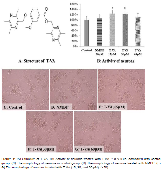
As shown in B, the control group was set as 100 % neuronal activity based on the optical density (OD) values of the MTT assay. T-VA promoted neuron activity in all treatment groups, but the doses of 15 and 30 μM showed more significant effect (p < 0.05) than the high dose of 60 μM. The activity of neurons in NMDP group also increased compared with the control group. Under optical microscopy, neurons in control group exhibited pyramidal cell body with fine dendritic networks (C). More neurons are evident in the NMDP group compared with control group (D). In addition, the number of neurons and the dendritic networks increased with various concentrations of T-VA. Notably, the T-VA doses of 15 and 30 μM induced better neuron growth than the high dose (60 μM) group (Figures 1E-1G).
Effect of T-VA on differentiated PC12 cells
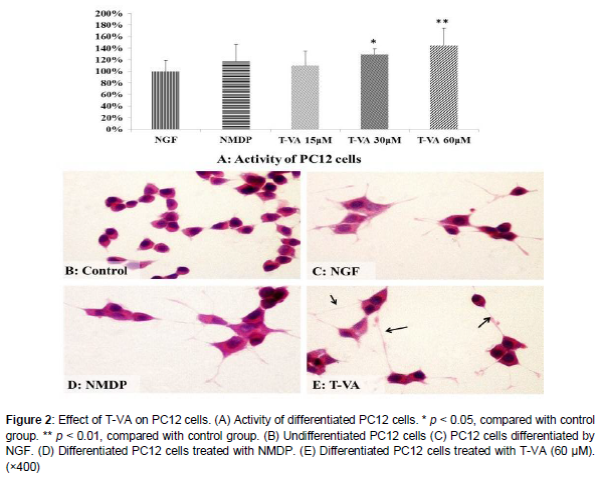
The NGF group was set as 100 % activity of PC12 cells. Results of the MTT assay showed that T-VA at 15, 30, and 60 µM increased cell viability to 110.14 %, 128.99 % and 144.93 %, respectively in a dose-dependent manner. Observation of morphologic changes of PC12 cells revealed that undifferentiated PC12 cells proliferated to form clone-like cell clusters without neural characteristics (B), that PC12 cells differentiated by NGF had round cell bodies with fine dendritic networks and intact and clear cell edges (C), and that cells in NMDP group had long synapse (D). PC12 cells treated with T-VA had the longest synapse of all the groups and an obvious increase in the amount of neurite-bearing cells compared with the control group (E).
Expression of VEGF in differentiated PC12 cells
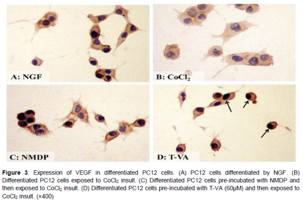
The immunohistochemical results of VEGF are represented in . The CoCl2 group showed weak expression of VEGF (B). The brown staining of the NGF and NMDP groups was obviously increased (Figures 3A and 3C). Moreover, the T-VA group (60 μM) displayed significant expression of VEGF compared with the CoCl2 group (D).
Microvascular proliferation of T-VA on CAM
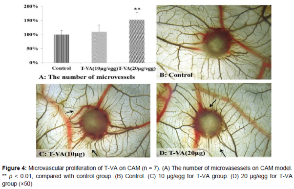
T-VA had microvascular proliferation effect on CAM. When the dose of T-VA was 10 μg/egg, the impact of promoting microvascular proliferation was not very obvious (Figures 4A and 4C). However, when the dose was increased to 20 μg/egg, microvascular proliferation increased significantly compared with control group (p < 0.01) (A). As exhibited in D, we found that the amount of microvascular distinctly increased compared with the control group.
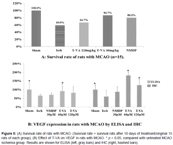
Effect of T-VA on survival rate and VEGF expression in rats with MCAO
Both survival rate and VEGF expression were markedly reduced in MCAO rats with ischemia compared with survival rate and VEGF expression in sham-operated rats, which were set as 100 % (Figures 5A and 5B). Survival rates in MCAO rats after treatment with 120 and 60 mg/kg of T-VA were increased by 6.7 and 26.7 %, respectively, compared with the survival rate in the untreated MCAO ischemia group. The survival rate in the MCAO rats after treatment with NMDP was also increased (by 20 %) compared with the survival rate in the untreated MCAO ischemia group. The areas of VEGF expression, determined by both ELISA and immunohistochemistry (IHC), were significantly decreased in the untreated MCAO ischemia group compared with VEGF expression in the sham-operated group (B).
The results of immunohistochemistry (IHC) are in agreement with the ELISA results. ELISA results revealed that the VEGF expression increased significantly in the T-VA (60 mg/kg) group (B) and IHC results revealed a significant increase in VEGF expression in both T-VA treatment groups (60 and 120 mg/kg) compared with that in the untreated MCAO ischemic group. The contents of VEGF in the 60 and 120 mg/kg T-VA treatment groups were 25.67 and 22.86 ng/mg protein, respectively, which corresponds to a 26.26 % and 16.28 % increase, respectively, compared with the VEGF content in the untreated MCAO ischemia group (18.28 ng/mg protein). NMDP treatment did not significantly improve the VEGF expression in rats with MCAO.
Discussion
It was previously reported that T-VA has a protective effect against injured PC12 cells (EC50 = 4.249 µM), whereas the congener structures of T-VA possess protective effects against damaged vascular endothelial cells [17-19]. In the present study, T-VA displayed the ability to promote the activities of neurons and PC12 cells (p < 0.05), and histopathological analyses coincided with the cell viability detected by the MTT assay. Overall, T-VA exhibited a positive effect on neurons in this study.
VEGF, a major mediator of angiogenesis, plays an important role in recovery of ischemic injury and serves as a useful positive control [34,35]. Our present study showed that T-VA exhibited the ability to stimulate VEGF expression in PC12 cells. Further study using CAM model showed that T-VA significantly promoted micro-angiogenesis (p < 0.01). These findings suggest that there might be a correlation between VEGF expression and cell viability. When the rats were treated with T-VA, VEGF up-regulated dramatically, thereby preventing MCAO rats from ischemic injury. Additionally, this study indicates that the survival rate of MCAO rats after treatment with T-VA was 26.67 % higher than that in the ischemia group, suggesting an effective approach against ischemic injury.
Obviously, it is of vital importance to increase the survival rate for the patients. All the findings in present study suggest that the neuroprotective effect of T-VA may be related to an increase of VEGF expression, laying the foundation for further research of the mechanism. This study also indicates the feasibility of discovering more efficient compounds from traditional Chinese medicine via structure combination.
Conclusion
T-VA promotes the activities of neurons and differentiated PC12 cells and increases the expression of VEGF both in vitro and in vivo in rats. Therefore, T-VA may be a potential candidate in preventing cerebral ischemic injury.
Declarations
Acknowledgement
References
Archives


News Updates